The Perils of Alcohol Consumption for Fatty Liver Patients

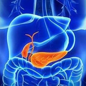
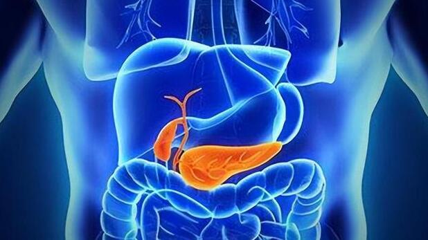
The discovery of fatty liver during a physical examination often goes unnoticed by many due to the lack of overt symptoms. This complacency, however, can be detrimental as even mild cases of fatty liver disease can impair liver function. Recent studies have indicated that female patients with fatty liver disease may face greater risks compared to their male counterparts.

A study published in the Journal of Hepatology has revealed that among patients with alcohol-related fatty liver disease, women have a 1.6 times higher mortality risk within a certain period compared to male patients.

In this study, Metabolic and Alcohol-Related Fatty Liver Disease (MetALD) is defined as the consumption of 20-50 grams of alcohol per day (or 140-350 grams per week) for women, and 30-60 grams per day (or 210-420 grams per week) for men, exhibiting characteristics of metabolic dysfunction-associated fatty liver disease.

The study included over 10,000 participants, among whom 1,971 suffered from various types of fatty liver disease. Although the incidence rate of fatty liver disease in men was about twice that of women, over a span of 26.7 years, women exhibited a significantly higher mortality risk.

Compared to individuals without liver disease, female MetALD patients had an 83% higher risk of all-cause mortality than their male counterparts; and when compared to male patients with alcohol-related liver disease, the mortality risk for women was 160% higher.

This disparity is attributed to the lower levels of alcohol dehydrogenase in the stomach lining of women, which results in higher blood ethanol levels in women after consuming the same amount of alcohol. This leads to more intact alcohol reaching the liver, triggering or exacerbating the progression of fatty liver disease.

Additionally, alcohol can weaken the intestinal barrier function, leading to dysbiosis. When this occurs, intestinal bacteria can transfer endotoxins to the liver. Based on body weight, the level and duration of endotoxins in women are higher than in men after consuming corresponding amounts of alcohol.

Dietary Choices Matter for Fatty Liver Patients

For those with fatty liver disease, it is crucial to control calorie intake while maintaining a balanced diet. Here’s how to approach different types of food:

Meat and Eggs

Diets should ideally include a moderate amount of lean meat, eggs, and fish (a total of 150 grams per day), which provide essential nutrients such as high-quality protein, lecithin, and vitamins.

Consume less offal and avoid high-fat, high-cholesterol foods like animal organs, chicken skin, pig brain, etc. Also, limit consumption of meat broth, chicken soup, and fish soup.

Vegetables

Ensure a daily intake of 500 grams of vegetables. The following vegetables are recommended:

Celery contains dietary fiber, vitamins, minerals, etc., which help reduce fat and cholesterol.

Kelp is rich in taurine, which can lower cholesterol in bile.

Tomatoes have a beneficial health effect on fatty liver and can be eaten raw or cooked.

Onions not only have antibacterial properties but also help regulate blood lipids.

Garlic helps reduce cholesterol in the blood and prevent thrombosis.

Main Foods

Strictly control the intake of staple foods, up to a maximum of half a kilogram per day, especially reducing the proportion of refined grains such as white rice, white porridge, white bread, etc.

Consume more coarse grains, which contain more minerals, vitamins, and dietary fiber. For example, oats are rich in linoleic acid and saponins, helping to reduce serum cholesterol and triglycerides.

Fruits

Fruits contain a wealth of minerals and vitamins, as well as various beneficial active substances. It is recommended to consume 200-350 grams of fruit daily.

Citrus fruits are rich in vitamins and aid in fat metabolism, increasing the liver’s metabolic detoxification rate. Fatty liver patients can consume them regularly. High-calorie and high-sugar fruits should be consumed in moderation, such as durian, sugarcane, raisins, etc.

Beverages

Soy products like soy milk are low in energy and are recommended to be consumed at least once a day; milk contains more calcium, which can inhibit the activity of cholesterol synthase in the human body, and it is advisable to choose low-fat milk.

Sugars in drinks and various sweets can easily be converted into fat in the liver and should be avoided as much as possible. It is best not to drink alcohol.

Fats

Eat less or avoid high-fat content foods such as fried foods. Animal fats like lard, butter, cream, etc., which are high in saturated fatty acids, should be avoided, and daily vegetable oil intake should be 20-25 grams.

Starting with the Details to Detoxify the Liver

The clinical manifestations of fatty liver are diverse, with mild fatty liver often showing no clinical symptoms and usually discovered incidentally during physical examinations.

Fatigue is the most common symptom reported by patients with fatty liver. Moderate to severe fatty liver can present similarly to chronic hepatitis, with symptoms such as loss of appetite, fatigue, nausea, vomiting, and vague pain in the liver area or upper right abdomen. Unhealthy lifestyle habits are the main risk factors leading to fatty liver, and making a few changes in life can help detoxify the liver.

Reject All Alcohol

Resolutely avoid all alcoholic beverages. With a damaged liver, alcohol cannot be fully broken down and remains in the body over time, accelerating the progression of fatty liver. Women, especially those with a family history or related risk factors, need to be particularly strict with their alcohol intake.

Gradually Control Weight

Overweight or obese patients with fatty liver should focus on weight loss. A study published in the journal Gastroenterology showed that if weight is reduced by more than 5%, symptoms improve in 58% of patients with fatty liver. It is recommended to lose 0.5-1 kilogram per week and reduce body weight by more than 5% within a year until a healthy weight is achieved.

Create a Caloric Deficit

Ensure a balanced intake of fats, proteins, vitamins, and trace elements, with a daily energy intake in negative balance by 500-1000 kilocalories to create a caloric deficit. The Mediterranean diet can help reduce liver fat content, with increased consumption of vegetables, fruits, fish, whole grains, beans, and olive oil.

Maintain Regular Exercise

Regular exercise, such as aerobic, resistance, and flexibility training, can help reduce intrahepatic lipids and improve liver function.

It is recommended to engage in aerobic exercise for more than 30 minutes but not exceeding one hour each time, at least five times a week, with a maximum heart rate during exercise of (170 – age) beats per minute. Options include swimming, jogging, cycling, brisk walking, dancing, etc.

For those with poor cardiopulmonary function or who are intolerant to aerobic exercise, resistance training can be chosen, performing light to moderate resistance muscle training 2-3 times a week, such as lifting dumbbells, push-ups, resistance band training, etc.

Related Posts

Comprehensive Understanding of Gallbladder Cancer: Risks, Symptoms, Diagnosis, and Prevention Strategies

Gallbladder cancer, though not the most common type of cancer, poses a significant threat to health due to its stealthy symptoms and the prevalence of late-stage diagnoses. As a malignant tumor in the digestive system, it primarily originates from the epithelial cells of the gallbladder mucosa. Confronting this challenge requires us to identify its causes, stay alert to its potential symptoms, and employ a range of diagnostic methods for early detection, while adopting effective preventive measures to reduce its occurrence.

Risk Factors for Gallbladder Cancer

The etiology of gallbladder cancer is linked with several risk factors:

  1. Gallstones: Chronic presence of gallstones is a significant factor, with 70%-90% of patients having a history of cholelithiasis.
  2. Chronic Cholecystitis: Recurrent inflammation of the gallbladder can potentially lead to cancer.
  3. Gallbladder Polyps: Particularly those exceeding 1 centimeter in diameter.
  4. Family History: Some families exhibit a higher prevalence of the disease.
  5. Age and Gender: The elderly and women are more susceptible.
  6. Obesity: A known risk factor for many types of cancer, including gallbladder cancer.

Symptoms of Gallbladder Cancer

Early stages of gallbladder cancer are often asymptomatic. However, as the tumor grows and spreads, signs may include:

  1. Upper Abdominal Pain: Possibly persistent or intermittent.
  2. Jaundice: A yellowing of the skin and eyes typically due to bile duct obstruction by the tumor.
  3. Unexplained Weight Loss: A rapid decrease in weight without an obvious cause.
  4. Loss of Appetite: Potentially accompanied by nausea and vomiting.
  5. Abdominal Mass: Sometimes palpable through touch.

While these symptoms can be disconcerting, timely detection and proactive treatment can tip the scales in favor of conquering the disease.

Diagnosing Gallbladder Cancer

Diagnosis of gallbladder cancer involves:

  1. History and Physical Examination: Inquiry into symptoms and history of gallstones or other biliary diseases, along with a physical search for abnormalities like abdominal masses or ascites.
  2. Blood Tests: Liver function tests (e.g., ALT, AST, ALP, total and direct bilirubin) to reflect the status of the liver and biliary system. Tumor markers like CA19-9 and CEA may also be elevated, although non-specifically.
  3. Imaging Studies: Ultrasound is often the initial imaging modality, revealing wall thickness, internal structure, and tumor presence. CT scans offer detailed images of the gallbladder and surrounding organs, aiding in assessing tumor size, location, and metastasis. MRI provides extensive information on soft tissue structures, particularly useful for biliary changes. ERCP and EUS with close examination of the biliary tree can detect minute alterations.
  4. Histopathological Examination: If imaging suggests a tumor, tissue samples obtained through surgery or biopsy are examined pathologically to confirm cancer. Pathology is the “gold standard” for diagnosing gallbladder cancer.
  5. Additional Tests: PET-CT may be used to evaluate cancer spread.

Comprehensive diagnosis of gallbladder cancer necessitates integrating clinical presentations, laboratory findings, and imaging studies, ultimately confirmed by pathological examination. Early diagnosis and treatment are pivotal for improved prognosis. Please consult medical professionals for specific medical advice, as the information provided is for educational purposes only.

Treatment of Gallbladder Cancer

Treatment is closely related to the individual’s circumstances, with the most effective options being:

  1. Surgical Removal: If the tumor has not spread, surgery to remove the gallbladder and surrounding tissue can be curative.
  2. Chemotherapy and Radiation: For inoperable patients, these modalities can control disease progression and extend survival.

Surgery is the only potentially curative approach for gallbladder cancer. However, given the poor prognosis and low long-term survival rates, early screening and diagnosis are critical.

Preventing Gallbladder Cancer

While there are no absolute rules for preventing gallbladder cancer, adopting a healthy lifestyle may reduce the risk:

  1. Maintain a Healthy Weight: Obesity is a risk factor for various cancers, including gallbladder cancer. Balanced diet and regular exercise help maintain a healthy weight.
  2. Healthy Dietary Habits: High-fiber diet with ample fruits, vegetables, and whole grains; limit high-fat and high-cholesterol foods; reduce processed foods and red meat intake.
  3. Control Gallstones: Since they are a major risk factor, maintaining a healthy weight and dietary habits can help control their formation.
  4. Quit Smoking: Smoking is linked to many cancers, and quitting can lower the risks, possibly including that of gallbladder cancer.
  5. Regular Medical Check-ups: If you have conditions that increase the risk of gallbladder cancer (such as gallstones or chronic cholecystitis), regular check-ups can help in early detection and appropriate interventions.
  6. Avoid Rapid Weight Loss and Yo-yo Dieting: Extreme dieting methods or frequent weight fluctuations may increase the risk of gallstones, a risk factor for gallbladder cancer.
  7. Caution with Hormone Replacement Therapy: Some studies suggest HRT may increase the risk of gallbladder cancer. Women should discuss the pros and cons with their doctors before considering these therapies.
  8. Genetic Counseling: If gallbladder cancer runs in the family, consider genetic counseling to understand your risk and get professional advice.

Though there’s no foolproof method to completely prevent gallbladder cancer, these measures may help lower the risk.

In conclusion, we must emphasize that gallbladder cancer is not insurmountable. With proactive engagement, early detection, and vigorous treatment, victory over this disease is within reach. Additionally, we must attend to our emotional well-being and mindset, maintaining a cheerful disposition and fostering confidence in overcoming illness.

Let us all be vigilant about gallbladder cancer, elevate our awareness, strengthen self-care, and take responsibility for our health.

Mastering Diabetes Management: A Comprehensive Guide to Home Care and Self-Management

Diabetes, a chronic condition often underestimated in its severity, can significantly impact not only the patient but also every family member when it becomes an unwelcome guest in the household. The importance of home care for diabetes patients cannot be overstated, as it plays a crucial role in managing the condition and improving the patient’s quality of life. Here’s how to effectively manage diabetes at home:

The Importance of Family Care for Diabetes Patients

Managing diabetes requires long-term treatment and care, with the home environment being crucial for disease control and enhancing the patient’s quality of life. Good family care helps stabilize conditions, improves the patient’s lifestyle, and reduces the financial burden on the family.

1. Healthy Eating: Fueling the Body

The Art of Balanced Food Intake

  • Prioritize Low GI Foods: Opting for foods with a low glycemic index, such as whole grains and legumes, helps stabilize blood sugar levels.
  • A Feast of Colorful Vegetables: Rich in fiber, vitamins, and minerals, a colorful plate not only looks appealing but is also beneficial for health.
  • Precise Carbohydrate Portions: Diabetes patients don’t need to avoid carbs entirely but should learn to calculate and control their intake.

Personalized Diet Plans

  • As each patient’s situation is unique, collaborate with a professional dietitian to create a personalized meal plan based on weight, blood sugar levels, and activity levels.

2. Exercise Therapy: The Natural Foe of Diabetes

Gradual and Daily Exercise

  • Regularity: Choose suitable exercises and ensure at least 150 minutes of moderate-intensity activity per week.
  • Variety: Combine cardio and strength training to better control blood sugar by engaging more muscle groups.

Monitoring and Adaptation

  • Monitor blood sugar before and after exercise to understand the impact and adjust diet and insulin dosage accordingly.

3. Blood Sugar Management: The Secrets Behind the Numbers

Precision Monitoring

  • Regular Testing: Use a glucometer regularly to monitor blood sugar levels and record data to identify patterns.
  • Smart Technology: Utilize Continuous Glucose Monitoring (CGM) systems for real-time glucose insights and timely adjustments.

Responding to Highs and Lows

  • Low Blood Sugar: Keep fast-acting sugar sources like glucose tablets or juice on hand.
  • High Blood Sugar: Follow medical advice to adjust diet and medication promptly.

4. Psychological Support: Dancing with the “Sweet” Disease

  • Positive Communication: Family members should listen to patients’ feelings and face the challenges together.
  • Professional Assistance: Seek help from mental health experts for counseling or therapy if needed.

5. Regular Check-ups: Better Safe Than Sorry

  • Routine Examinations: Regularly check blood pressure, lipids, eye exams, and foot inspections to detect and treat issues early.
  • Self-Examination: Teach patients how to self-check, especially feet, daily for wounds or abnormalities.

6. Medication Treatment: Vital Pill-Taking Discipline

  • Medication Adherence: Take hypoglycemic and related medications strictly as prescribed.
  • Drug Knowledge: Understand the mechanisms of action, potential side effects, and how medication relates to diet and exercise.

Considerations for Family Caregivers

Regular Blood Sugar Monitoring: Help patients monitor blood sugar regularly and seek medical attention for significant fluctuations or persistent increases.

Complication Prevention: Be aware of common complications like cardiovascular disease, retinopathy, and diabetic nephropathy. Encourage healthy habits like quitting smoking and maintaining adequate sleep to reduce risks.

Health Education: Participate in diabetes education to enhance care skills and teach patients self-care techniques like correct blood sugar measurement and insulin injections.

Through these home care strategies, families can help patients stabilize blood sugar, reduce complication risks, and improve their quality of life. The essence of diabetes home care lies in integrating knowledge with action, establishing good habits, and ensuring a supportive family environment. With proper self-management skills and a positive outlook, patients can navigate their daily lives with confidence, maintain good blood sugar control, and delay complications, sharing a fulfilling life with their loved ones.

Herpes Zoster: Understanding, Prevention, and Treatment Strategies

Herpes Zoster, commonly known as shingles or zoster, is an acute infectious disease caused by the Varicella-zoster virus (VZV), the same virus responsible for chickenpox. After the initial infection manifests as chickenpox, the virus doesn’t leave the body but lies dormant in the nerve ganglia. Upon weakening of the immune system, the virus can reactivate and cause a painful rash along the distribution of the affected nerve, leading to the characteristic symptoms of shingles.

Triggers that Awaken the Volcano: The Onset of Shingles

What could awaken this dormant “volcano” within us? The primary factors contributing to this reactivation are:

  1. Immune System Weakening: Age-related decline, high stress, lack of rest, or other illnesses can impair our immune response.
  2. Other Influences: Certain medications, particularly those that suppress the immune system like anti-rejection drugs used after organ transplants, can trigger the reactivation of the virus.

Signs of Viral Reawakening: Symptoms of Herpes Zoster

Shingles does not appear silently; it often announces its arrival with the following signals:

  1. Pain That Precedes the Rash: Often serving as a harbinger of the disease, this pain can start days before the appearance of the rash.
  2. A Chain of Skin Volcanoes: Subsequently, small blisters filled with fluid develop on one side of the body, sitting on a red base.
  3. Persistent Burning and Stinging: The affected area becomes sensitive, with even light touch causing discomfort.
  4. Additional Symptoms Alongside the Rash: These may include fever, headache, and fatigue.

Treatment: Strategies to Quell the “Volcano”

Once shingles erupt, prompt treatment is crucial:

  1. Antiviral Therapy:
    Initiating antiviral medication early (usually recommended within 72 hours after the rash appears) with drugs like acyclovir, valacyclovir, or famciclovir can halt further viral replication, shortening the episode and reducing severity.
  2. Pain Management:
    Targeted pain relief can be achieved with over-the-counter medications like acetaminophen or ibuprofen, as well as prescription-strength gabapentin, pregabalin, or amitriptyline for managing acute nerve pain. For severe pain, a short course of low-to-medium dose corticosteroids (like prednisone) may be necessary to reduce inflammation and nerve pain.
  3. Local Care:
    Keeping the affected area clean and dry prevents secondary infections from broken blisters. Topical treatments like acyclovir cream can help suppress viral activity. If blisters have ruptured, antibiotic ointments may prevent bacterial infections, with cold or wet compresses providing relief from pain and itching.
  4. Physical Therapy:
    Depending on the condition, physical therapy methods such as heat therapy, laser treatment, or ultraviolet light exposure can accelerate healing and alleviate pain.
  5. Neurotrophic Medications:
    Supplements like the B-vitamin group and cobalamin aid in nerve repair and can prevent or reduce postherpetic neuralgia.

It’s worth noting that each individual’s response to treatment varies, and a personalized treatment plan should be crafted based on the patient’s specific needs and the severity of their condition. It’s important to seek medical attention as soon as possible, as early antiviral treatment yields better outcomes.

Preventive Measures: A Fire Alert for Shingles

  1. Vaccination Against Chickenpox: The chickenpox vaccine can reduce the risk of both chickenpox and shingles. Consult a healthcare provider for vaccination schedules suitable for you.
  2. Strengthening Immunity: Maintaining healthy habits that enhance immune function includes adequate sleep, a balanced diet, moderate exercise, and stress reduction.
  3. Avoid Direct Contact with Shingles Patients: Since shingles are contagious through the virus, avoiding close contact with someone who has it reduces the chance of transmission.
  4. Good Personal Hygiene: Regular handwashing, especially after touching potentially contaminated objects or surfaces, helps prevent viral entry through the eyes, nose, or mouth.
  5. Preventing Viral Reactivation: For those who have had chickenpox, shingles result from viral reactivation. Maintaining good health and avoiding prolonged stress can help prevent this.

If you suspect you have contracted shingles, seek immediate medical advice for accurate diagnosis and appropriate treatment recommendations.

While shingles are not incurable, the pain they cause should not be underestimated. Prevention is always better than cure. Cultivating healthy living habits, monitoring your immune status, and getting vaccinated when appropriate are powerful tools in our fight against shingles.

Esophageal Cancer: Understanding, Prevention, and Treatment Strategies

In the silent corners of our daily lives, a silent and lethal disease could be lurking within our thrice-daily meals – Esophageal Cancer. Globally, hundreds of thousands of people succumb to this disease annually. In China, the incidence and mortality rates of esophageal cancer are particularly high, making it a significant health threat.

Esophageal cancer is a malignant tumor that occurs in the esophagus, the tube that transports food from the throat to the stomach. The main types include Squamous Cell Carcinoma and Adenocarcinoma.

Signs of Esophageal Cancer: Symptoms Not to Be Overlooked

Initially, esophageal cancer may not exhibit obvious symptoms. However, as the condition progresses, warning signals may appear, including:

  1. Difficulty Swallowing: This is usually the first symptom to appear, initially manifesting as trouble swallowing dry, hard, or large foods.
  2. Sore Throat or Pain Behind the Chest Bone
  3. Weight Loss and Fatigue
  4. Coughing or Vomiting: Some patients may vomit blood or have black stool, signs of internal bleeding.
  5. Hoarseness If you experience any of these symptoms, especially persistent difficulty swallowing, seek medical attention immediately.

The Secret of Esophageal Cancer: Uncontrolled Growth of Mutated Cells

Esophageal cancer is a malignant tumor formed by uncontrolled proliferation of cells on the inner lining of the esophagus. It mainly has two types:

  1. Squamous Cell Carcinoma: Originates from the squamous cells lining the esophagus and is closely linked to smoking and drinking.
  2. Adenocarcinoma: Common near the stomach part close to the esophagus and related to Acid Reflux and Barrett’s Esophagus (a condition associated with long-term acid reflux).

Causes and Risk Factors: A Multifactorial Symphony

The specific causes of esophageal cancer are not fully understood, but certain factors significantly increase the risk:

  1. Smoking and Excessive Alcohol Consumption: Both can damage esophageal cells and increase the likelihood of mutations.
  2. Poor Diet: A diet lacking fruits and vegetables may raise the risk of esophageal cancer.
  3. Gastroesophageal Reflux Disease (GERD): Long-term acid reflux can damage the esophageal mucosa.
  4. Barrett’s Esophagus: An esophageal change caused by GERD, posing a higher risk.
  5. Obesity: Increases the severity of gastroesophageal reflux.
  6. Family History and Genetic Factors Moreover, long-term consumption of high-fat, high-salt, and barbecued foods may also increase the incidence of esophageal cancer.

Diagnosis: Key Steps Unveiling the Truth

  1. Clinical Symptoms and History: Doctors will thoroughly understand symptoms like dysphagia, chest pain, weight loss, etc., and ask about high-risk factors such as smoking, drinking history, and family history of esophageal cancer patients.
  2. Endoscopy: Upper gastrointestinal endoscopy is the most common and crucial method for diagnosing esophageal cancer. If suspicious abnormalities are found, doctors may take tissue samples for histopathological examination (biopsy), which is the gold standard for diagnosis.
  3. Imaging Studies: Include chest X-rays, CT scans, MRI, PET-CT, etc., to assess tumor location, size, invasion depth, and metastasis.
  4. Endoscopic Ultrasound (EUS): More accurately detects the depth of tumor invasion into the esophageal wall and surrounding lymph node status.
  5. Esophagography: By swallowing barium and undergoing X-ray fluoroscopy, it clearly shows changes in the shape of the esophagus, helping to identify stenosis, obstruction, and other lesions.
  6. Blood Tests: Although not directly diagnostic for esophageal cancer, abnormal levels of tumor markers like CEA and CA19-9 in the blood may suggest the possibility of cancer.

A definitive diagnosis of esophageal cancer requires comprehensive information from various sources. Once suspected, visit a qualified hospital promptly for an individualized treatment plan based on the doctor’s assessment.

Treatment: The Arsenal Against Cancer

  1. Surgery:
    Classic open surgeries include left-sided thoracotomy (Ivor-Lewis surgery), right-sided thoracotomy, and transthoracic combined-approach surgery, aimed at completely removing the cancerous part of the esophagus and reconstructing esophageal function through procedures like esophagogastric anastomosis or esophageal replacement surgery (like colonic interposition).
    In recent years, minimally invasive surgeries like total thoracoscopic and laparoscopic esophagectomy have rapidly developed, completing surgery through small incisions, reducing trauma and accelerating recovery.
  2. Radiation Therapy:
    Mainly used for patients unsuitable for surgery or at high surgical risk, especially those with upper esophageal cancer or locally advanced esophageal cancer. Radiation can be used alone or combined with chemotherapy (concurrent chemoradiation) to shrink tumors, control disease progression, and improve quality of life.
    Advanced techniques like Intensity-Modulated Radiation Therapy (IMRT), Image-Guided Radiation Therapy (IGRT), and Stereotactic Body Radiation Therapy (SBRT) have improved radiation therapy effectiveness and safety.
  3. Chemotherapy:
    Chemotherapy can be used as neoadjuvant therapy to shrink tumors before surgery, as adjuvant therapy to reduce recurrence after surgery, and as palliative treatment for advanced or metastatic esophageal cancer.
    New chemotherapy drugs and targeted drug applications, along with the rise of immunotherapy, have provided more treatment options for esophageal cancer patients.
  4. Endoscopic Therapy:
    For early-stage esophageal cancer (carcinoma in situ or intramucosal cancer), endoscopic mucosal resection (EMR) or endoscopic submucosal dissection (ESD) can be performed.
    For mid-to-late stage patients unable to undergo surgery or before surgery, endoscopic placement of stents, laser thermotherapy, photodynamic therapy, etc., can relieve obstructive symptoms and improve eating difficulties.
  5. Comprehensive Treatment:

    The treatment of esophageal cancer increasingly leans towards multimodal comprehensive treatment, such as preoperative neoadjuvant chemoradiotherapy + surgery, postoperative adjuvant chemoradiotherapy, etc., and a reasonable combination of various therapies to maximize treatment effects, reduce side effects, and extend patient survival.
    In conclusion, the treatment of esophageal cancer is individualized and should be assessed and recommended by clinicians to develop the most suitable treatment plan for the patient. Additionally, supportive treatment during the recovery phase, nutritional support, and psychological counseling are also essential components of the overall treatment plan.

Prevention: Proactive Lifestyle Changes

While there is no definitive way to completely prevent esophageal cancer, the following lifestyle adjustments can significantly reduce risk:

  1. Lifestyle Related:
    Quitting Smoking: One of the primary causes of esophageal cancer, avoiding smoking can significantly lower the risk of developing the disease.
    Limiting Alcohol Consumption: Excessive alcohol consumption is also linked to esophageal cancer, so controlling or avoiding alcohol altogether is recommended.
    Maintaining a Healthy Weight: Being overweight or obese increases the risk of many cancers, including esophageal cancer. A healthy diet and exercise plan should be adopted to maintain a healthy weight.
    Avoiding Long-Term Exposure to Hazardous Substances: Some occupational exposures increase the risk of esophageal cancer, such as long-term exposure to asbestos, benzene, etc. Ensure proper protective measures at work.
  2. Dietary Habits:
    Consuming Plenty of Fruits and Vegetables: These contain fiber, vitamins, and trace elements that can reduce the risk of esophageal cancer.
    Reducing Processed Meat and Red Meat Intake: Excessive consumption of processed and red meat may increase the risk of certain cancers; it’s advisable to substitute with fish, poultry, etc.
    Avoiding High-Temperature Foods: Regularly consuming very hot foods and beverages may damage the esophageal mucosa and increase the risk of cancer.
    Avoiding Food Contamination: Some foods may contain carcinogens like aflatoxins; ensure proper storage and handling to prevent mold contamination.
  3. Medical Monitoring and Intervention:
    Monitoring Barrett’s Esophagus: Those with GERD that progresses to Barrett’s esophagus are at higher risk and require regular endoscopy and biopsy.
    Treating GERD Promptly: Effectively treating GERD can reduce its progression to Barrett’s esophagus and even esophageal cancer.
    Screening for High-Risk Groups: Individuals with a family history of esophageal cancer or other high-risk factors may require more frequent screening.
    Preventing esophageal cancer involves improving living habits, adjusting diet, and medical monitoring. If any physical discomfort or specific risk factors exist, consult a doctor promptly for guidance and treatment.

Understanding esophageal cancer is crucial for preventing and treating this disease. Through public education, we can raise awareness of esophageal cancer, enhance prevention consciousness, and lower incidence rates. At the same time, knowledge helps patients better understand their condition, cooperate with treatment, and improve recovery rates.

While esophageal cancer is frightening, it is not invincible. As long as we understand its causes, preventive measures, and treatment methods, actively adjust our lifestyle, maintain a positive mindset, we can effectively reduce the risk of developing this disease and conquer it.

Spring’s Unwelcome Guest: The Guide to Triumphing Over Urticaria

Spring, the season of rejuvenation and blossoming beauty, brings with it not only the promise of new life but also a common, yet unwelcome guest – urticaria, or what we commonly refer to as hives. This seemingly sudden skin reaction is particularly prevalent in the warm embrace of spring, silently intruding on our lives during an otherwise enchanting season.

What exactly is urticaria? At its core, urticaria represents a localized swelling response caused by the dilation and increased permeability of small blood vessels in the skin and mucous membranes. It manifests as sudden patches of redness or wheals on the skin, often accompanied by intense itching.

The Typical Culprits of Spring Urticaria:

Pollen and Catkins – The Invisible Challengers: During spring, pollen and catkins become ubiquitous allergens. As these minute particles drift through the air, they can easily be inhaled or settle on the skin, triggering an immune response that leads to urticaria.

Fluctuating Temperatures – An Unstable Instigator: The erratic temperature shifts characteristic of spring can provoke abnormal vasodilation and constriction responses in the skin, potentially eliciting hives.

Clothing Friction – An Unexpected Catalyst: Lightweight spring attire and the friction from changing clothes can exacerbate sensitive skin, especially for those predisposed to allergies.

Symptoms to Watch For:

Skin Erythema: Usually sudden in appearance, these are clear-bordered elevations varying in shape and size.
Itching: Often, the erythema is accompanied by significant itchiness.
Shape-Shifting: The patches may change shape or location over time, sometimes within minutes or hours.
Edema: Slight swelling may occur around the erythematous areas.
Transience: Each patch typically subsides within 24 hours, though new ones may appear elsewhere.
Pressure Sensitivity: Applying pressure can turn the erythema white (known as “blanching urticaria”).
Mild Pain or Stinging: Some patients may experience slight stinging or pain.
Urticaria can be acute or chronic. Acute episodes resolve within hours to weeks, typically lasting up to six weeks. Chronic urticaria persists beyond six weeks, with symptoms possibly recurring continuously. If hives are accompanied by difficulty breathing, throat swelling, or other systemic symptoms, it could indicate an anaphylactic emergency requiring immediate medical attention.

How to Fortify Against Spring Urticaria:

  1. Avoid Known Allergens: If certain substances are known to trigger your allergies, minimize contact with them.
  2. Limit Outdoor Activities: Reduce outdoor activities during peak pollen times, especially early morning and evening.
  3. Dress Appropriately: Wear long sleeves to reduce direct contact with allergens.
  4. Maintain Indoor Cleanliness: Regularly clean floors and surfaces, use air purifiers, and keep windows closed to reduce indoor pollen and dust mites.
  5. Hygiene Rituals: Shower and change clothes immediately upon returning home to remove adhered allergens.
  6. Balanced Diet: A healthy diet can strengthen the immune system’s defenses.
  7. Avoid Irritants: Steer clear of cosmetics with strong fragrances or certain laundry detergents that might irritate the skin.
  8. Stress Management: Implement stress reduction techniques, as stress can exacerbate urticaria symptoms.
  9. Medication Awareness: Be mindful of drugs like NSAIDs that could trigger hives and consult a doctor if a connection is suspected.
  10. Regular Monitoring: Those with chronic urticaria should report symptoms regularly to their physician for treatment adjustments.

If hives frequently occur without a clear allergen identified, professional medical assistance is advisable. Allergy testing may help determine specific triggers for a targeted prevention plan.

When urticaria symptoms arise, prompt medical attention is crucial. Doctors may prescribe antihistamines or topical antipruritics to manage symptoms.

Coping with Urticaria: What You Need to Know…

Upon developing hives, avoid panic. To alleviate symptoms, consider the following:

  1. Cold Compression: Using a cool towel or ice pack can ease itching and reduce swelling.
  2. Medication Use: Over-the-counter antihistamines like cetirizine can provide relief. Seek medical advice for severe symptoms.
  3. Psychological Adjustment: Stress management is key, as stress can trigger urticaria.
  4. Medical Consultation: If self-care fails, seek dermatological assistance promptly.

While spring urticaria can be vexing, understanding and employing preventive and responsive strategies significantly mitigate its impact on our lives. Let’s embrace the splendor of spring without the disturbance of hives. Remember, a healthy lifestyle and a positive outlook are potent remedies against any adversity.

Stroke and Paralysis: A Journey Towards Resilience and Recovery

Stroke, a term that may seem distant yet can strike at any moment, has brought untold pain and turmoil to many families. Paralysis, one of the common aftermaths of a stroke, often leaves patients and their loved ones feeling helpless. However, this is not the end of hope, but rather the beginning of a journey to reclaim control over one’s life.

What is a Stroke?

A stroke, medically referred to as a cerebrovascular accident, occurs when there is a blockage (ischemic stroke) or rupture (hemorrhagic stroke) in the blood vessels supplying the brain, leading to a shortage of blood and subsequent neurological dysfunction. When a stroke affects the part of the brain controlling one side of the body, it results in the loss of motor and sensory functions on that side – a condition known as hemiplegia due to stroke.

When Stroke Meets Paralysis

Hemiplegia signifies the loss of normal motor ability on either the left or right half of the body. This symptom indicates that the damage is not just muscular but extends to the neural pathways in the brain that control limb movement.

Unveiling the Story Behind Strokes and Paralysis

In the event of a stroke, time is synonymous with the life of brain cells. Approximately 1.9 million neurons perish with each passing minute due to ischemia. As time elapses, the surviving neural networks diminish, leading to a reduction or even complete loss of muscle control.

Symptoms of Post-Stroke Paralysis

  1. Limb weakness or paralysis: Patients may experience weakness or an inability to move one side of the body, including the face, arm, and/or leg.
  2. Facial droop: A stroke can cause one side of the facial muscles to malfunction, resulting in asymmetrical facial expressions, particularly noticeable when smiling.
  3. Speech difficulties: Damage to the speech center can lead to slurred speech and reduced ability to understand or express language.
  4. Visual impairments: A stroke can impair vision in one or both eyes, causing partial blindness or blurriness.
  5. Coordination disorders: Difficulties in walking, loss of balance, dizziness, or challenges in coordinated movements may arise.
  6. Sensation abnormalities: One side of the body may experience tingling, numbness, or unusual sensations.
  7. Cognitive dysfunction: Decreased memory, concentration difficulties, and executive function impairments may occur.
  8. Emotional fluctuations: Post-stroke patients may experience mood swings, such as irritability, depression, or symptoms of depressive disorders. These symptoms can occur suddenly and may be temporary or permanent. If any of these symptoms arise, immediate medical attention should be sought, as a stroke is a medical emergency. Prompt treatment is crucial for minimizing brain damage and enhancing recovery prospects.

Prevention is More Important Than Treatment

  1. Healthy diet: Consume plenty of fruits and vegetables, reduce salt and oil intake to maintain unobstructed blood vessels.
  2. Regular exercise: Moderate physical activity can strengthen heart function and promote blood circulation.
  3. Weight control: Being overweight or obese is a risk factor for stroke.
  4. Quit smoking and limit alcohol: Nicotine in cigarettes can cause blood vessels to constrict, while alcohol may lead to increased blood pressure.
  5. Regular check-ups: Timely detection and treatment of underlying conditions such as hypertension and diabetes are essential.

What to Do When a Stroke Occurs?

>If you suspect yourself or someone else is having a stroke, immediately dial emergency services. Remember the “FAST” principle:

F (Face): Is there a drooping side of the face?
A (Arms): Is one arm unable to lift?
S (Speech): Is the speech slurred or unclear?
T (Time): Time is brain cells; act now!

Recovering from Paralysis – Not a Fairy Tale, But Science

In the recovery process post-stroke, patients need to relearn how to use affected body parts through physical and occupational therapy. It’s a challenging journey, but science has shown that the brain is malleable and can reorganize itself to compensate for the damage caused by a stroke.

  1. Treating Paralysis: A Multifaceted Approach
  2. Physical therapy: Professional therapists can help patients with targeted training to gradually regain muscle strength and coordination.
  3. Occupational therapy: Training in daily living skills helps patients adapt to life and improve their quality of life.
  4. Medication: Certain drugs can help improve blood circulation and reduce spasticity.
  5. Surgery: In some cases, surgery may be used to remove clogged vessels or repair ruptured ones.
  6. Speech therapy: If speech abilities are impaired, speech therapy is also essential.

Beyond medical treatments, the patient’s own efforts and persistence are key to overcoming post-stroke paralysis. Through active rehabilitation training, patients can gradually enhance their quality of life and regain confidence in living.

On the road to recovery from paralysis, patients require courage, perseverance, and support. Although the journey may not be smooth sailing, every small step forward is worth celebrating. The support of family, friends, and the medical team is an indispensable force on this journey.

Post-stroke paralysis is not the end of life but an opportunity to redefine oneself. Despite the challenges ahead, scientific and medical advancements have made stroke recovery more than a mere fantasy.